
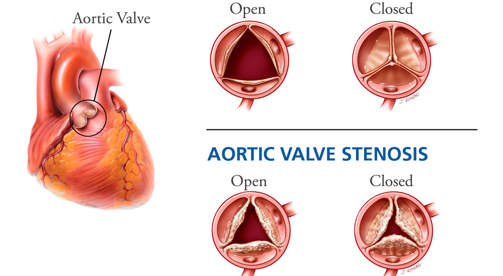
The aorta is the largest blood vessel in the body and distributes blood throughout it. The aortic valve is located between the left ventricle and the main artery that carries blood to the body (the aorta). When the heart contracts, the aortic valve opens and oxygen-rich blood is ejected into the aorta. Narrowing (stenosis) of the aortic valve makes it difficult for the heart to pump blood to the body.
In short, aortic valve stenosis is a condition that causes complications by obstructing the outflow of blood from the left ventricle; it occurs in 1–2% of people over 65.
Causes: rheumatic disease, calcific degeneration, congenital bicuspid valve
A healthy aortic valve has three leaflets. In congenital aortic stenosis, instead of three leaflets the valve has one thick, stiff leaflet or two leaflets. Sometimes the narrowing may be in the tissue below the valve (subaortic stenosis), or in the aorta itself just above the valve.
The main diagnostic method is echocardiography. Catheterization is needed in patients for whom surgery is planned. The basic principle of treatment is to relieve the narrowing; for this purpose, valve replacement with a prosthetic valve and balloon valvuloplasty are performed. In high-risk patients, transfemoral aortic valve implantation (TAVI) or balloon valvuloplasty is used.
Information on this site is general and does not replace an examination by a doctor.